Laminitis in donkeys (Astrid Arnold)
Why we should not compare horse and donkey laminitis. It is generally known that donkeys are not horses with long ears. Donkeys have their own feeding and husbandry requirements, and own particular social behaviour. Even so, we still tend to treat donkey hooves the same way as we treat horse hooves. However, donkeys are quite different from horses – not only in their requirements on husbandry, but also in their lower limb anatomy.
A donkey may have a coffin bone, navicular bone, pastern, fetlock and so on, but there are still major differences in the bone texture, shape, aspect ratios and structures involved. Tendons and ligaments in donkeys are far more flexible than they are in horses. Passively moving a donkey’s leg – especially the carpal joint – will reveal an astonishing range of movement.
A donkey’s hoof capsule is different from that of a horse in both shape and structure. The toe walls are very thick in donkeys while the side walls without the bevelling are relatively thin. The heels are also shaped differently. The coffin bone on a donkey has no palmar or plantar processes, so the heel fulfills that function. Even the horn has a different quality – donkey hooves swell up faster and to a far greater extent in moist surroundings.
There are also differences in blood circulation – the blood vessels are injected with synthetic resin during specimen preparation. Donkey blood vessels take around 8 to 10 ml of resin compared to only about 5 to 6 ml in comparable ponies while preparing the specimen, even though donkey legs are slenderer.
1 Comparison in hoof and coffin bone anatomy
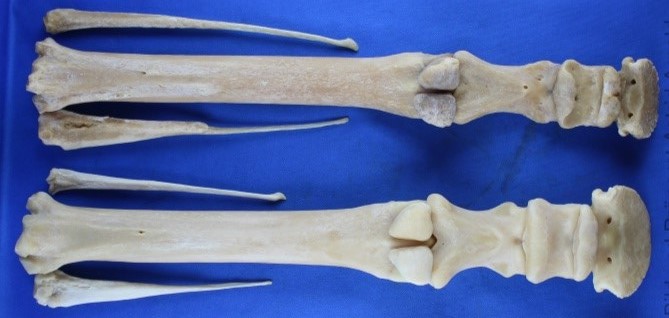
Figure 1: Hind legs – donkey above, pony below.
Donkey bones are generally slenderer and longer in proportion to circumference.

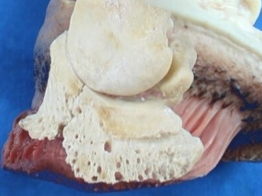
Figure 2: Hind hooves – pony left, donkey right. The most striking difference is in the coffin bone, which is considerably smaller, lower, and without plantar or palmar processes in donkeys. This gives the collateral cartilage less support from the coffin bone than in horses.

Figure 3: Hind hooves – pony left, donkey right. The donkey’s coffin bone may be smaller, but the horn capsule is around the same size as that of the pony shown for comparison. Both animals were around the same size with around the same leg length up to the hock.


Differences from a horse’s hoof must be taken into account in order to make any sense evaluating X-ray images from a donkey. In particular, coffin bone density in a donkey cannot be compared with a horse.
There are hardly any X-rays of healthy donkey legs, so I x-rayed Oskar, a young and healthy donkey, some years ago. Oskars X-rays have since saved many other donkey lives. Changes in the coffin bone in donkeys with laminitis may appear dramatic at times, but they seem far less serious compared to those of a healthy donkey rather than a healthy horse.
I believe it is extremely important to draw attention to the differences in the coffin bone to prevent any more donkeys from being put down due to diagnostic error.
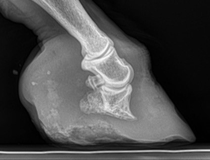
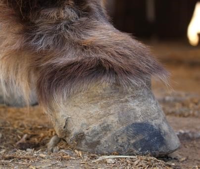
Figures 4 to 6: Front left hoof of the young and healthy donkey Oskar


Figures 7 to 9: X-ray images of the donkey Oskar, front left, Courtesy of Dr. Feigl


Figures 10 to 12: X-ray images of a horse for comparison, Courtesy of Dr. Marcus Menzel


2 Tendons and ligaments
Another significant difference between donkeys and horses lies in tendon and ligament flexibility, which is far more pronounced in donkeys. This explains the relatively high likelihood of dislocation and subluxation issues in donkeys. On the other hand, donkeys suffer far less frequently from clubfoot than horses do – neglected donkey hooves in particular are far more likely to develop sinking pasterns.
3 The hoof capsule
Figures 13 and 14: Sagittal section of donkey hoof, lateral and medial


The cartilage in horses is largely supported by the plantar and palmar processes that are missing in donkey hooves; donkey cartilage is mainly supported on the heel as Figure 13 clearly shows.
As Figure 3 clearly shows, the horn walls on the toe are far thicker in donkeys; donkey hooves have fewer cornified lamellae, which do however have a sturdier shape. The donkey hoof’s sole is very thick, at least at the forehand; this makes the front of the hoof very stable, and the toe wall is less likely to deform due to laminitis compared to a horse.
4 Laminitis in donkeys
Apart from characteristic horn fold formation, donkey hoof capsules show very late changes such as the toe wall beginning to protrude with laminitis. This kind of change requires a very high and long toe wall in a donkey.
Even so, internal changes will take place just as fast as in horses. This applies not only to a widening suspensory apparatus due to toe wall leverage that always takes priority in horses – even without visible toe wall levering, the corium may change with atrophying or lipping in the coffin bone. A donkey may still have a perfectly decent horn capsule even with these changes. Internal changes are hardly noticeable from outside, with a loose wall in the toe area as the only change noticed.
Figures 15 to 17: Specimen preparation of a donkey front hoof with laminitis
5 Emergency call: Felix the donkey
I received an emergency call in July 2016 – a local animal protection organisation had rescued two donkeys from inappropriate husbandry.
One case required immediate trimming. I had already met these two donkeys in 2006 for trimming. Felix had pronounced changes in his hooves, indicating laminitis.
I could not achieve any improvement in husbandry, feeding or regular trimming for the donkeys at the time, and we gradually lost contact. I was supposed to see the donkey after ten years in 2016, at which time they probably already had chronic untreated laminitis. My prognoses were therefore poor.

Figure 19 shows Felix’s front hooves while he was still in the stable from where he was taken from. No photos of the soles were made, and Felix was barely able to stand up. I trimmed his hooves as an emergency measure on the weekend he was taken, and Felix was more able to stand up although walking was very difficult for him, which was also partly because he was emaciated and weak. It is easy to imagine how steep the coffin bone must have been from the visibly upwardly compressed ungular cartilage.


After two weeks, X-rays were taken towards a more accurate assessment of the situation (Figures 20 – 21). Note that the pastern joint, not the coffin joint, was now at the same height as the coronet – yes, the coffin bone had sunk that far down into the hoof capsule.
Felix was lame on his poorer front left-hand hoof at the time. It quickly turned out that the lameness was due to a hoof ulcer in the loose wall.
Figures 20 and 21: X-ray front right and left lateral in July 2016, Courtesy of Dr. Marcus Menzel


Figures 22 and 23: Felix’s hooves front right and left at the time of X-ray in July 2016

Felix’s coffin bones were assessed as highly atrophied, and he had also been lying down for long periods. The veterinary authority and a number of vets recommended euthanasia. Again, a donkey hoof was compared to a horse and assessed as more problematic than it actually was for the donkey.
However, the X-rays from Oskar the donkey showed the necessity of assessing a donkey differently, and how it made sense to wait and watch Felix’s progress once the hoof ulcer had been treated and left time to heal.
Figure 24, 25: X-ray front right and left lateral in March 2017, Courtesy of Sabine Wallner, vet

Figures 26 and 27: Felix’s hooves front right and left at the time of X-ray in March 2017
We can never expect these hooves to return to the anatomy we would like to see. The coffin bone sunk down to the pastern joint will remain that way. Any change in hoof position will affect Felix’s short pastern bone as well as the coffin bone, like these two bones were one. Felix is only mobile from the pastern joint upwards. Finding the most functional hoof angle for Felix is still a challenge.
Felix likes to join his donkey friends for a walk nowadays.