Previous medical history
Paula's suffering started in the summer of 2008. An X-ray of her right forehoof showed that she had a hollow hoof wall and that her coffin bone was rotated. This diagnosis led to Paula's first hoof wall resection.
After being treated for a year it was possible to ride her again. It lasted until the April of 2010 when again the farrier found her hoof wall was hollow, but this time it was her left forehoof that was affected. This led to another resection of the hoof wall and a subsequent shoeing.
In the September of 2010 Paula again went lame. The vet X-rayed her left forehoof and diagnosed a coffin bone rotation, which led to another hoof wall resection and treatment with EquipalazoneR. He remarked that every other warmblood would have already been put down. So Paula should be thankful for „only“ being a Haflinger. (shows Paula's luck to be „only“ a Haflinger.)


Alternative treatment methods
October 1st 2010: Her owner decided to stop the treatment with EquipalazoneR and had Paulas shoes removed. She was looking for an alternative treatment. A barehoof practicioner advised to bring Paula to a specific kind of equine hoof clinic. There she was told not to visit within the first six months, because most owners can't bear to see the pain their horses are in. But the treatment was said to be very important to the healing process. Paula's owner refused and kept looking for another solution. She found the forum of DHGeV and described the devastating state her horse was in. November 21st 2010: Paula gets her first orthopaedic hoof treatment.


Coffin bone rotation
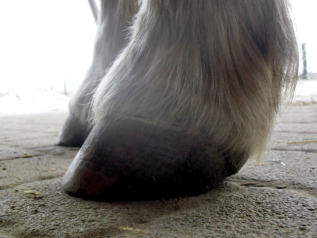
The long and slanted toe wall of her right forehoof, resected in 2008, shows the problems of the regrown hoof wall , due to the laming and resection history. A new hollow wall is just about to build up. Looking from the sole view you are already able to see the torn connection of sole and wall. Putred horn tubules indicate another hollow wall and coffin bone rotation. In reality Paula’s coffin bone hadn’t moved at all, as the diagnosis coffin bone rotation would suggest. It actually was the toe wall which shifted in relation to the coffin bone.
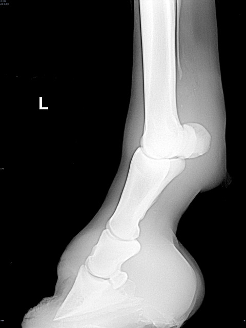
The x-rays of both hooves show straight bone columns. The coffin bone hasn’t rotated in both hooves. The rotting and levering toe wall of the left fore hoof had been removed 14 days prior.
A sensible hoof treatment should prevent and eliminate cause of such a gap between toe wall and coffin bone. This means that the toe wall has to be handled in such a way that it can keep itself short and that there can be no manual trimming of Paula’s heels.


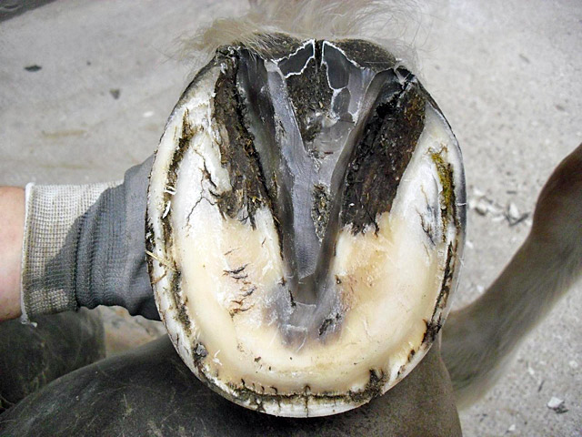
The toe wall has grown down all the way. From the sole view you see that the hoof platelets still suffer the consequences of last years damage. A toe wall resection, as Paula had to cope with several times, creates a great danger to the very sensitive dermis of the hoof. By removing the protective layer above the coffin bone carrier, the laminar horn loses moisture and gets hard and brittle. The hoof platelets which are tightly connected to the laminar horn suffer the most. It results in a vulnerable white line. The laminar layers of both hooves improve during the following months due to the orthopaedic treatment. But they never reach a realy healthy state again. Paula will probably keep this scar.
She was able to be ridden two months after the first orthopaedic hoof treatment. On trail tracks shes wearing boots. Even though Paula never had laminitis, her owner always keeps care of enough movement and suitable food management. (green guard muzzle), so that Paula stays in shape. Paula's hooves are effectively being cared for in variety of ways.